

Nervous system
Objectives
At the end of this
lecture, student will be able to
• List the structures of nervous system
• Explain various functions of nervous system
• Describe the basic histology of nervous tissue
• Differentiate between various types of neurons
• Explain the neurohumoral transmission of nervous system
• Describe the role of neurotransmitter in the neurohumoral
transmission
• Explain the organisation of nervous system
• Distinguish the physiology of various nervous systems
• List the various parts of brain
• Describe the protective coverings of brain
• Explain the importance of blood brain barrier
• Describe the role of cerebrospinal fluid
• List the various parts of brain stem
• Describe the anatomy of brain stem
• Explain the physiology and functions of various parts of
brain stem
• Describe the anatomy of cerebellum
• Explain the functions of cerebellum
• List the various components of diencephalon
• Describe the physiology and functions of components of
diencephalon
• Describe the role of hypothalamus
• Describe the functions of circumventricular organs
• Explain the structure and function of cerebrum
• List and locate the lobes of the cerebrum
• Describe the nuclei that comprise the basal ganglia
• List the structures of the limbic system
• Describe the functions of limbic system
• Explain the locations and functions of various areas of
cerebral cortex
• Define “brain waves”
• Indicate the significance of brain waves
• List various cranial nerves
• Identify the cranial nerves by name, number and type
• Describe the location and functions of cranial nerves
• Describe the protective structures of spinal cord
• Describe the gross anatomical features of the spinal cord
• Explain spinal nerves connected to the spinal cord
• Describe the functions of the major sensory and motor
tracts of the spinal cord
• Describe the functional components of a reflex arc and the
ways reflexes maintain homeostasis
• Describe pre ganglionic and post ganglionic neurons of the
autonomic nervous system
• Compare the anatomical components of the sympathetic and
parasympathetic divisions of the autonomic nervous system
• Describe the components of an autonomic reflex
• Explain the relationship of the hypothalamus to the ANS
Content
−Structures of nervous system
−Functions of nervous system
−Histology of nervous tissue
−Types of neurons
•Neurohumoral transmission
•Organization of nervous system
•Physiology of nervous systems
•Parts of brain
•Protective coverings of brain
•Blood brain barrier
•Cerebrospinal fluid
Anatomy and Physiology of
– Brain stem
– Cerebellum
– Diencephalon
– Hypothalamus
– Circumventricular organs
– Cerebrum
– Basal ganglia
– Limbic system
• Physiology of cerebral cortex
• Brain waves
Cranial nerves
– Types
– Location
– Functions
• Spinal cord
– Protective structures
– Anatomical features
– Sesory and motor tract
• Spinal nerves
• Reflex arc
• Autonomic nervous system
– Sympathetic
– Parasympathetic
• Autonomic reflex
• Hypothalamus and ANS
Nervous
System
• Mass of 2 kg, 3% of total body
• One of the smallest and yet the most complex systems
• The structures include:
– Brain
– Cranial nerves and their branches
– Spinal cord
– Spinal nerves and their branches
– Ganglia
– Enteric plexuses
– Sensory receptors

• The skull encloses the brain – contains about 100 billion
neurons
• 12 pairs cranial nerves, numbered I - XII
• Emerge from the base of the brain
• Nerve:
– Bundle of axons + associated connective tissue & blood
vessels
– Lies outside the brain and spinal cord
– Follows a defined path
– Serves a specific region of the body

Cranial Nerves
Name of cranial nerves
• 0lfactory nerve
• Optic nerve
• Oculomotor
nerve
• Trochlear nerve
• Trigeminal
nerve
• Abducent nerve
• Facial nerve
•
Vestibulocochlear nerve
•
Glossopharyngeal nerve
• Vagus nerve
• Accessory nerve
• Hypoglossal
nerve
• The Spinal Cord
– Connects to the brain through the foramen magnum of the
skull
– Encircled by the bones of the vertebral column
– Contains about 100 million neurons
– 31 pairs of spinal nerves emerge from the spinal cord

Spinal Nerve
• 31 pairs
• Spinal nerve follows the name of corresponding vertebra
column.
• Consists cervical spinal nerve, thoracic spinal nerve,
lumbar spinal nerve, sacral spinal nerve and coccyx spinal nerve.
• Emerge from spinal cord and through the intervertebral
foramina of vertebra.
• Spinal nerves:
1. 8 pairs of
cervical spinal nerves
2. 12 pairs of
thoracic spinal nerves
3. 5 pairs of
lumbar spinal nerves.
4. 5 pairs of
sacral spinal nerves
5. 1 pairs of
coccyx spinal nerves.

• Ganglia
– Consisting primarily of neuron cell bodies
– Located outside the brain and spinal cord

Functions
of Nervous System
• Carries out a complex array of tasks
• Allows us to sense various smells
• Produce speech
• Remember past events
• Provides signals that control body movements
• Regulates the operation of internal organs
• These diverse activities can be grouped into three basic
functions:
– Sensory
– Integrative
– Motor
Sensory function
• Sensory receptors detect internal or external stimuli
• Carried into the brain and spinal cord through cranial and
spinal nerves

• Sensory receptor
– Dendrites of sensory neurons
– Specialized cells that monitor changes in the internal or
external environment

Integrative function
• Processes sensory information by analyzing and storing
some of it and by making decisions
• Perception - the conscious awareness of sensory stimuli
Motor function
• Elicit an appropriate motor response by activating
effectors through cranial and spinal nerves
• Causes muscles to contract and glands to secrete
Histology
of Nervous Tissue
• Neurons:
– Provide most of the unique functions of the nervous system
• Neuroglia:
– Support, nourish, protect the neurons and maintain
homeostasis
– Smaller than neurons, 5 to 50 times more numerous
– Do not generate or propagate action potentials

The cell body
(Perikaryon or soma)
• Contains a nucleus & typical cellular organelles:
lysosomes, mitochondria, and a golgi complex and free ribosomes
• Prominent clusters of rough ER (nissl bodies)
• The cytoskeleton: Neurofibrils & microtubules
• Lipofuscin - Product of neuronal lysosomes

Nerve Fiber
• Neuronal process (extension) emerges from the cell body of
a neuron
• 2 kinds of processes: multiple dendrites and a single axon

• Dendrites (little
trees)
– The receiving or input portions of a neuron
– Short, tapering, and highly branched
– Cytoplasm contains Nissl bodies, mitochondria, and other
organelles
• Axon
– Propagates nerve impulses toward another neuron, a muscle
fiber, or a gland cell
– Long, thin, cylindrical projection that often joins the
cell body
– At a cone-shaped elevation called the axon hillock
– Closest to the axon hillock is the initial segment
• The cytoplasm of an axon, called axoplasm
• Surrounded by a plasma membrane known as the axolemma
• Side branches - axon
collaterals
• The axon and its collaterals end by dividing into many
fine processes called axon terminals

Structural
Classification of Neurons

Functional
classification of Neurons
• Motor or efferent neurons
• Sensory or afferent neurons
• Interneuron or association neurons

Neuroglial
Cells
• Astrocytes –
Cover capillaries of brain to form BBB and help regulate passage of molecules
from blood to brain
• Oligodendrocytes –
Form myelin sheath around central axons producing the white matter of CNS

• Microglia –
Phagocytic amoeboid cells in CNS, remove foreign and degenerated material from
brain
• Ependymal cells -
Line the ventricles or brain cavities and central canal of spinal cord
• Schwann cells -
Surround axons of all peripheral nerve fibres, form the myelin sheath
• Satellite cells -
Supply nutrients to the surrounding neurons and also have some structural
function, protective, cushioning cells

Myelination
• Multilayered lipid and protein covering
• Electrically insulates the axon
• Increases the speed of nerve impulse conduction
• Two types of neuroglia produce myelin sheaths:
– Schwann cells (in the PNS)
– Oligodendrocytes (in the CNS)
• Axons without such a covering – unmyelinated

Collections
of nervous tissue
• Nerve
– Bundle of axons
– Located in the PNS
– Cranial nerves connect the brain to the periphery
– Spinal nerves connect the spinal cord to the periphery
• Tract
– A bundle of axons
– Located in the CNS
– Interconnect neurons in the spinal cord and brain

• White matter
– Composed primarily of myelinated axons
– Appear whitish
• Gray matter
– Neuronal cell bodies, dendrites, unmyelinated axons, axon
terminals & neuroglia
– Appears grayish; Nissl bodies impart a gray color

Neurohumoral
Transmission
• Plasma
membranes of presynaptic and postsynaptic neurons - separated by the synaptic
cleft
• Nerve impulses
cannot conduct across the synaptic cleft
• In response to
a nerve impulse, the presynaptic neuron releases a neurotransmitter
• Diffuses
through the fluid in the synaptic cleft binds to receptors in the plasma
membrane of the postsynaptic neuron
• The
postsynaptic neuron receives the chemical signal produces a postsynaptic
potential
Synapse

Role of Pre
and Post synaptic Neurons
• Thus, the
presynaptic neuron converts an lectrical signal (nerve impulse) into a chemical
signal (released neurotransmitter)
• The
postsynaptic neuron receives the chemical signal and in turn generates an
electrical signal (postsynaptic potential)
Neurohumoral
Transmission

• A nerve impulse
arrives at a synaptic end bulb of a presynaptic axon
• The
depolarizing phase of the nerve impulse opens voltage gated ca channels
• Calcium ions
are more concentrated in the EC fluid - flows inward through the opened
channels
• Increase in the
concentration of ca - triggers exocytosis of the synaptic vesicles
• As vesicle
membranes merge with the plasma membrane, neurotransmitter released into the
synaptic cleft
• Each synaptic
vesicle contains several thousand molecules of neurotransmitter
• Diffuse across
the synaptic cleft and bind to neurotransmitter receptors in the postsynaptic
neuron’s plasma membrane
• Binding of
neurotransmitter – opens the channels and allows particular ions to flow across
the membrane
• As ions flow
through the opened channels, the voltage across the membrane changes
• This change in
membrane voltage is a postsynaptic potential
• Postsynaptic
potential may be a depolarization or a hyperpolarization
• When a
depolarizing postsynaptic potential reaches threshold, it triggers an action
potential in the axon of the postsynaptic neuron
Excitatory
Postsynaptic Potentials
• A neurotransmitter
that depolarizes the postsynaptic membrane is excitatory
• A depolarizing postsynaptic potential - Excitatory postsynaptic potential (EPSP)
• Although a single EPSP normally does not initiate a nerve
impulse, the postsynaptic cell does become more excitable
Inhibitory
Postsynaptic Potentials
• A neurotransmitter that causes hyperpolarization of the
postsynaptic membrane is inhibitory
• During hyperpolarization, generation of an action
potential is more difficult
• Membrane potential becomes inside more negative
• Even farther from threshold than in its resting state
• A hyperpolarizing postsynaptic potential - an inhibitory post synaptic potential
(IPSP)
Neurotransmitter
Receptors
• Ionotropic receptor
• Metabotropic Receptor

Removal of
the Neurotransmitter
Diffusion
• Some diffuse away from the synaptic cleft
Enzymatic degradation
• Inactivated through enzymatic degradation
• Example - the enzyme acetylcholinesterase breaks down
acetylcholine in the synaptic cleft
Uptake by cells
• Many neurotransmitters are actively transported back into
the neuron that released them (reuptake)
• Others are transported into neighboring neuroglia (uptake)
Neurotransmitters
• Neurotransmitters can be divided into two classes based on
size: small-molecule neurotransmitters and neuropeptides
• The small-molecule
neurotransmitters include:
– Acetylcholine, amino acids, biogenic amines, ATP and other
purines, and nitric oxide
• Neuropeptides:
– Substance P, encephalin, endorphin and dynorphin
Organisation
of Nervous system

Organisation
of the CNS
• Consists of the brain and spinal cord
• Processes many different kinds of incoming sensory
information
• The source of thoughts, emotions, and memories
• Most nerve impulses that stimulate muscles to contract and
glands to secrete originate in the CNS

Organization
of the PNS
• Includes all nervous tissue outside the CNS
• Components of the PNS include:
– Cranial nerves and their branches
– Spinal nerves and their branches
– Ganglia
– Sensory receptors
• Subdivided into SNS, ANS & ENS

Somatic
Nervous System
Consists of:
Sensory neurons
• Convey information from somatic receptors in the head,
body wall, and limbs
• From receptors for the special senses of vision, hearing,
taste, and smell to the CNS
Motor neurons
• Conduct impulses from the CNS to skeletal muscles only
• Motor responses - voluntary

Autonomic
Nervous System
Sensory neurons | Motor neurons |
• Convey information from autonomic sensory receptors • Convey information from autonomic sensory receptors | • Located primarily in visceral organs to the CNS • Conduct nerve impulses from the CNS to smooth muscle, cardiac • Involuntary • Motor part - two branches • Parasympathetic division - “rest-and-digest” activities • Sympathetic division - “fight-or-flight” responses |
Enteric
Nervous System
• “Brain of the gut,” involuntary
• Sensory neurons
– Monitor chemical changes and stretching within GIT
• Motor neurons
– Govern:
•
Contraction of GI tract smooth muscle to propel food
•
Secretions of the GI tract organs such as gastric acid
•
Activity of GIT endocrine cells - secrete hormones

Action potential
• An action potential (AP) or impulse is a sequence of
rapidly occurring events that decrease and reverse the membrane potential and
then eventually restore it to the resting state
• Two main phases:
– A depolarizing phase
– A repolarizing phase


Brain
• The adult brain consists of four major parts:
– Brain stem
– Cerebellum
– Diencephalon
– Cerebrum

• Brain stem
– Continuous with the spinal cord
– Consists of the medulla oblongata, pons, and midbrain
• Cerebellum
– Posterior to the brain stem
• Diencephalon
– Superior to the brain stem
– Consists of the thalamus, hypothalamus, and epithalamus
• Cerebrum
– Largest part of the brain
– Supported on the diencephalon and brain stem

Protective
Coverings of the Brain
• The cranium and the cranial meninges surround and protect
the brain
• The cranial meninges
– Continuous with spinal meninges
– Outer dura mater
– Middle arachnoid mater
– Inner pia mater

Cranial meninges

Extensions
of the dura mater
• Separate parts of the brain:
• Falx cerebri
– Separates the two hemispheres (sides) of the cerebrum
• Falx cerebelli
– Separates the two hemispheres of the cerebellum
• Tentorium cerebelli
– Separates the cerebrum from the cerebellum


Blood flow
to Brain
• Blood flows to the brain - via the internal carotid and
vertebral arteries
• Internal jugular veins return blood from head to heart
• Consumes about 20% of oxygen & glucose used even at
rest
• Neurons synthesize ATP from glucose using oxygen
• When increases in a region of the brain, blood flow to
that area also increases
• Virtually no glucose is stored in the brain (supply of
glucose must be continuous)
Blood Brain
Barrier
• Protects brain cells from harmful substances and pathogens
• Prevents passage of many substances from blood into brain
tissue
• Consists mainly of tight junctions - seal together the
endothelial cells of brain capillaries

• Along with a thick basement membrane around the
capillaries
• The processes of many astrocytes, press up against the
capillaries
• Secrete chemicals - maintain the permeability
characteristics of the tight junctions
• A few water-soluble substances (glucose) cross BBB by
Active transport
• Others - creatinine, urea, and most ions, cross the BBB
very slowly
• Proteins and most antibiotic drugs—do not pass at all
• However, lipid-soluble substances, such as oxygen, carbon
dioxide, alcohol, and most anesthetic agents, easily cross
Cerebrospinal
Fluid
• Clear, colorless liquid
• Protects the brain and spinal cord from chemical and
physical injuries
• Carries oxygen, glucose, and other needed chemicals from
the blood to neurons and neuroglia
• CSF continuously circulates through cavities in the brain
and spinal cord and in the subarachnoid space
• The total volume of CSF is 80 to 150 ml
• CSF contains glucose, proteins, lactic acid, urea, cations
(na, K, ca2, mg2), and anions (cl and HCO3)
• Also contains some WBC
Role of CSF
• The CSF contributes to homeostasis in three main ways:
Mechanical protection
• Shock-absorbing medium; protects the delicate tissues from
jolts; buoys the brain so that it “floats” in the cranial cavity
Chemical protection
• Provides an optimal chemical environment for accurate
neuronal signaling
• Even slight changes - seriously disrupt production AP
Circulation
• CSF allows exchange of nutrients and waste products
between the blood and nervous tissue
Ventricles
• CSF - filled cavities within the brain
A lateral ventricle
• Located in each hemisphere of the cerebrum
• Anteriorly, the lateral ventricles are separated by a thin
membrane - Septum pellucidum
The third ventricle
• Narrow cavity along the midline
• Superior to the hypothalamus
• Between the right and left halves of the thalamus
The fourth ventricle
• Lies between the brain stem and the cerebellum
• Contains some white blood cells
Ventricles of Brain

Formation
of CSF in the Ventricles
• The sites of CSF production are the choroid plexuses -
networks of blood capillaries in the walls of the ventricles
• Ependymal cells covering the capillaries form CSF from
blood plasma by filtration and secretion
• Ependymal cells are joined by tight junctions
• This blood–CSF barrier permits certain substances to enter
the CSF but excludes others
• Thus, protects the brain and spinal cord from potentially
harmful blood borne substances
Circulation
of CSF
• The CSF formed in the choroid plexuses of each lateral
ventricle
• Flows into the third ventricle through two narrow, oval
openings, the interventricular foramina
• The fluid then flows through the aqueduct of the midbrain
(cerebral aqueduct)
• Passes through the midbrain, into the fourth ventricle
• The choroid plexus of the fourth ventricle contributes
more fluid
• CSF enters the subarachnoid space through three openings
in the roof of the fourth ventricle: a median aperture and the paired lateral
apertures, one on each side
• CSF then circulates in the central canal of the spinal
cord and in the subarachnoid space around the surface of the brain and spinal
cord
CSF Flow

Choroid
plexus

Ventricles
of brain and flow of CSF

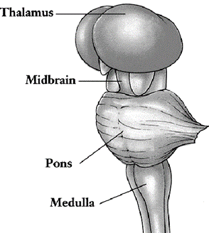
Brain Stem
• Part of the brain between the spinal cord and the
diencephalon
• It consists of three structures:
– Medulla oblongata
– Pons
– Midbrain
Reticular formation
• Extends through the brain stem Netlike region of
interspersed gray and white matter

Medulla
oblongata (Medulla)
• Continuous with spinal cord
• Forms the inferior part of brain stem
• Begins at the foramen magnum
• Extends to the inferior border of the po
• The medulla’s white matter contains all sensory
(ascending) tracts and motor (descending)
• Anterior aspect of the medulla has - pyramids

Medulla
oblongata - Pyramids
• Pyramids - Large corticospinal tracts, pass from the
cerebrum to the spinal cord
• Control voluntary movements of the limbs and trunk
• Just superior to the junction of the medulla with the
spinal cord – decussation of pyramids

Medullary
nuclei
• The medulla also contains several nuclei
• Some of these nuclei control vital body functions
• Cardiovascular
center
– Regulates the rate and force of the heartbeat and the
diameter of blood vessels
• The medullary
rhythmicity area - respiratory center
– Adjusts the basic rhythm of breathing

Medulla also control reflexes for:
• The vomiting center
• The deglutition center
• Sneezing
• Coughing
• Hiccupping
Olive
• Just lateral to each pyramid is an oval-shaped swelling
called an olive
Inferior olivary
nucleus

• Receives input from the cerebral cortex red nucleus of the
midbrain, and spinal cord
• Regulate the activity of cerebellar neurons
• Provides instructions that the cerebellum uses to make
adjustments to muscle activity as you learn new motor skills
Left
gracile nucleus and cuneate nucleus
• Nuclei associated with sensations of touch, pressure,
vibration, and conscious proprioception
• Located in the posterior part of the medulla

Nuclei of
sensory pathways
Medulla also contains nuclei - components of sensory
pathways for: gustation (taste), audition (hearing) & equilibrium (balance)
The gustatory nucleus
• Part of the gustatory pathway from the tongue to the brain
The cochlear nuclei
• Part of the auditory pathway from the inner ear to the
brain
The vestibular nuclei
• Components of the equilibrium pathway from the inner ear
to the brain
Medullary
nuclei – Associated to Cranial Nerves
• Medulla contains nuclei associated with five pairs of
cranial nerves:
– Vestibulocochlear (VIII) nerves
– Glossopharyngeal (IX) nerves
– Vagus (X) nerves
– Accessory (XI) nerves (cranial portion)
– Hypoglossal (XII) nerves
Pons
• Lies superior to the medulla and anterior to the
cerebellum
• Is a bridge that connects different parts of the brain
with one another
• Connections are provided by bundles of axons
• Consists of nuclei, sensory tracts, and motor tracts

• Along with the medulla, the pons contains vestibular
nuclei that are components of the equilibrium pathway
• Other nuclei in the pons: The pneumotaxic area and the
apneustic area of the respiratory center

• Contains nuclei associated with the following four pairs
of cranial nerves:
– Trigeminal (V) nerves
– Abducens (VI) nerves

– Facial (VII)
nerves– Vestibulocochlear (VIII) nerves
Midbrain
(Mesencephalon)
• Extends from the pons to the diencephalon
• The midbrain contains both nuclei and tracts
• The anterior part contains paired bundles of axons –
cerebral peduncles
• The cerebral peduncles consist of axons of:
– Corticospinal
– Corticopontine
– Corticobulbar tracts
• Conduct nerve impulses from motor areas in the cerebral
cortex to the spinal cord, pons and medulla
Tectum
• The posterior part of the midbrain; Contains four rounded
elevations
Superior colliculi
(two superior elevations)
• Reflex centers for certain visual activities
• Visual stimuli elicit eye movements for tracking moving
images & scanning stationary images
• Reflexes for movements of the head, eyes, and trunk in
response to visual stimuli
Inferior colliculi
(two inferior elevations)
• Part of the auditory pathway
• Relay impulses from the receptors for hearing in the inner
ear to the brain
• Reflex centers for the startle reflex, sudden movements of
the head, eyes, and trunk
Reticular
Formation
• Brain stem consists of small clusters of neuronal cell
bodies (gray matter)
• Interspersed among small bundles of myelinated axons
(white matter)
• Exhibit a netlike arrangement is known as the reticular
formation

• Extends from the upper part of the spinal cord, throughout
the brain stem, and into the lower part of the diencephalon
• Neurons within the reticular formation have both ascending
(sensory) and descending (motor) functions
Reticular activating
system (RAS)
• Part of the reticular formation
• Consists of sensory axons that project to cerebral cortex
• Helps maintain consciousness and is active during
awakening from sleep
• Descending functions are to help regulate posture and
muscle tone, the slight degree of contraction in normal resting muscles
Cerebellum
• Occupies the inferior and posterior aspects of the cranial
cavity
• Has highly folded surface – greatly increases the surface
area
• Posterior to the medulla and pons
• Inferior to the posterior portion of the cerebrum

• A deep groove known as the transverse fissure, along with
the tentorium cerebelli
• Supports the posterior part of the cerebrum, separate the
cerebellum from the cerebrum

Cerebellum – Superior
or inferior view
• In superior or inferior views - shape of cerebellum
resembles a butterfly
• Central constricted area - vermis (Worm)
• The lateral “wings” or lobes - cerebellar hemispheres
• Each hemisphere consists of lobes separated by deep and
distinct fissures

Cerebellum
lobes
• The anterior lobe and posterior lobe govern subconscious
aspects of skeletal muscle movements
• The flocculonodular lobe on the inferior surface
contributes to equilibrium and balance

Cerebellar
cortex
• The superficial layer of the cerebellum
• Consists of gray matter in a series of slender
• Parallel folds called folia (leaves)
• Deep to the gray matter are tracts of white matter called
arbor vitae (tree of life) resemble branches of a tree
Cerebellar nuclei:
• Regions of gray matter; Deeper within the white matter
• Give rise to axons carrying impulses from the cerebellum
to other brain centers
Functions
of Cerebellum
Primary Function
• Evaluate how well movements initiated by motor areas in
the cerebrum are actually being carried out
• Detects the discrepancies in movements
• Sends feedback signals to motor areas of the cerebral
cortex, via thalamus
• Thus help correct the errors
• Smooth the movements
• Coordinate complex sequences of skeletal muscle
contractions
• Main brain region that regulates posture and balance
• Make possible all skilled muscular activities, from
catching a baseball to dancing to speaking
• May also have non motor functions such as cognition
(acquisition of knowledge) and language processing
Diencephalon
• Extends from the brain stem to the cerebrum
• Surrounds the third ventricle
• Includes: Thalamus, hypothalamus and epithalamus

Thalamus
• Consists of paired oval masses of gray matter organized
into nuclei with interspersed tracts of white matter
• Axons that connect thalamus and cerebral cortex pass
through internal capsule - thick band of white matter Lateral to thalamus

Internal Capsule

• Intermediate mass
Thalamus
(Interthalamic adhesion)
– A bridge of gray matter
– Joins the right and left halves of the thalamus
• Internal medullary
lamina
– A vertical y-shaped sheet of white matter
– Divides the gray matter of the right and left sides of the
thalamus
– Consists of myelinated axons - enter and leave the various
thalamic nuclei
Importance
of Thalamus
• Major relay station
– For most sensory impulses that reach the primary sensory
areas of the cerebral cortex from the spinal cord and brain stem
• Contributes to
motor functions
– By transmitting information from the cerebellum and basal
ganglia to the primary motor area of the cerebral cortex
• Role - maintenance
of consciousness
Thalamic
Nuclei

• Based on positions and functions
Anterior nucleus
• Input from the hypothalamus & sends output to the
limbic system
• Functions in emotions and memory
Medial nuclei
• Input from the limbic system and basal ganglia & send
output to the cerebral cortex
• Function in emotions, learning, memory, and cognition
Nuclei in the lateral
group
• Receive input from the limbic system, superior colliculi,
and cerebral cortex & send output to the cerebral cortex
• Dorsal nucleus functions in the expression of emotions
• Posterior nucleus and pulvinar nucleus help integrate sensory
information
Intralaminar nuclei
• Lie within the internal medullary lamina
• Make connections with the reticular formation, cerebellum,
basal ganglia & wide areas of the cerebral cortex
• Function in arousal (activation of the cerebral cortex)
and integration of sensory and motor information
Midline nucleus
• Forms a thin band adjacent to the third ventricle
• Function in memory and olfaction
Reticular nucleus
• Surrounds the lateral aspect of the thalamus, next to the
internal capsule
• Monitors, filters and integrates activities of other
thalamic nuclei
Ventral group
• Five nuclei are part of the ventral group
• Ventral anterior nucleus, ventral lateral nucleus, ventral
posterior nucleus, lateral geniculate nucleus, medial geniculate nucleus
Ventral
Group Nuclei
Ventral anterior
nucleus
• Receives input from the basal ganglia
• Sends output to motor areas of the cerebral cortex
• Plays a role in movement control
Ventral lateral
nucleus
• Receives input from the cerebellum and basal ganglia
• Sends output to motor areas of the cerebral cortex
• Plays a role in movement control
Lateral geniculate
nucleus
• Relays visual impulses for sight from the retina to the
primary visual area of the cerebral cortex
Medial geniculate
nucleus
• Relays auditory impulses for hearing from the ear to the
primary auditory area of the cerebral cortex
Ventral posterior
nucleus
• Relays impulses for somatic sensations from the face and
body to the cerebral cortex.
• Somatic sensations - touch, pressure, vibration, itch,
tickle temperature, pain & proprioception
Hypothalamus
• Small part of the diencephalon
• Located inferior to the thalamus
• Composed of a dozen or so nuclei in four major regions: – Mamillary,
Tuberal, Supraoptic & Preoptic
Hypothalamic
Regions

Mammillary region
• Adjacent to the midbrain
• Includes the mammillary bodies & posterior
hypothalamic nuclei
• Mammillary bodies - two, small, rounded projections that
serve as relay stations for reflexes related to the sense of smell
Tuberal region
• Widest part of the hypothalamus
• Includes dorsomedial, ventromedial & arcuate nucleus
• Stalk like infundibulum - connects the pituitary gland to
hypothalamus
• Median eminence - slightly raised region, encircles the
infundibulum
Supraoptic region
• Lies superior to the optic chiasm
• Contains paraventricular, supraoptic, anterior
hypothalamic & suprachiasmatic nucleus
Preoptic region
• Anterior to the supraoptic region
• Considered part of hypothalamus (regulates certain
autonomic activities)
• Contains the medial and lateral preoptic nuclei
Role of
Hypothalamus
• Controls many body activities
• Major regulators of homeostasis
• Monitor osmotic pressure, glucose level, certain hormone concentrations
& temperature of blood
• Has several very important connections with the pituitary
gland
• Produces a variety of hormones
• Some functions can be attributed to specific hypothalamic
nuclei
Functions
of Hypothalamus
Control of the ANS
• Controls and integrates activities of the ANS
• Example: Regulation of HR, movement of food through the
GIT & contraction of the urinary bladder
Regulation of
emotional and behavioral patterns
• Together with the limbic system
• Participates in expressions of:
– Rage, aggression, pain, pleasure & behavioral patterns
related to sexual arousal
Production of
hormones
• Releasing hormones and inhibiting hormones
• Released into capillary networks in the median eminence
• Bloodstream carries these hormones to anterior lobe of the
pituitary
• Cell bodies secretes two hormones (oxytocin, ADH)
• Transported to the posterior pituitary and release
Regulation of eating
and drinking
• Through feeding center, satiety center and thirst center
Control of body
temperature
• As body’s thermostat
• Directs the ANS to stimulate activities that promote heat
loss or production and retention
Regulation of
circadian rhythms
• Suprachiasmatic nucleus serves as the body’s internal
biological clock
• Establishes the circadian rhythms
• Receives input from the eyes (retina)
• Sends output to other hypothalamic nuclei, the reticular
formation, & pineal gland
Epithalamus
• Small region superior and posterior to the thalamus
• Consists of:
– Pineal gland
– Habenular nuclei

Pineal gland
• Size of a small pea
• Secretes the hormone melatonin
• Contribute to the setting of the body’s biological clock
• Controlled by the supra chiasmatic nucleus of hypothalamus
• Promote sleepiness
Habenular nuclei
• Involved in olfaction
• Emotional responses to odors

Circumventricular
organs (CVOs)
• Parts of the diencephalon
• Can monitor chemical changes in the blood (lack BBB)
• Include part of:
– Hypothalamus,
pineal gland, pituitary gland, and few other nearby structures
• Functionally:
– These regions coordinate homeostatic activities of the
endocrine and nervous systems
– Regulation of BP, fluid balance, hunger, and thirst
Cerebrum-
Seat of Intelligence
• Provides us with the ability to read, write & speak
• Make calculations and compose music
• To remember the past, plan for the future & imagine
things
• The cerebrum consists of:
– Outer cerebral cortex
– An internal region of cerebral white matter
– Gray matter nuclei deep within the white matter

Cerebral
cortex
• Region of gray matter
• Forms the outer rim of the cerebrum
• Contains billions of neurons
• Gyri or
convolutions - folds
• Fissures -
deepest grooves between folds
• Sulci -
shallower grooves between folds are
• Longitudinal
fissure - separates the cerebrum into cerebral
hemispheres

Corpus
callosum
• Cerebral hemispheres are connected internally by the
corpus callosum
• A broad band of white matter containing axons that extend
between the hemispheres

Lobes of the Cerebrum
• Each cerebral hemisphere subdivided into 4 lobes
• Named after the bones covering them:
– Frontal, Parietal, Temporal and Occipital lobes

• Central sulcus
– Separates the frontal lobe from the parietal lobe
• Lateral cerebral
sulcus (fissure)
– Separates the frontal lobe from the temporal lobe
• Parieto- occipital
sulcus
– Separates the parietal lobe from the occipital lobe
Cerebral
Lobes and Sulci

Cerebral
White Matter
• Consists primarily of myelinated axons in three types of
tracts
1. Association tracts
– Contain axons that conduct nerve impulses
– Between gyri in the same hemisphere

2. Commissural tracts
– Contain axons - conduct nerve impulses from gyri to other
(b/w hemisphere)
– Corpus callosum
– Anterior commissure
– Posterior commissure
3. Projection tracts
• Contain axons that conduct nerve impulses from the
cerebrum to lower parts of the CNS (vice versa)
• Example internal capsule contains both ascending and
descending axons
Basal
Ganglia
• Deep within each cerebral hemisphere are three nuclei
(masses of gray matter)
• Two are side-by-side, just lateral to the thalamus

Corpus
striatum
• Refers to the striated (striped) appearance. It includes:
• Lentiform nuclei
– Globus pallidus - closer to the thalamus
– Putamen - closer to the cerebral cortex
• Caudate nucleus
– Large head connected to smaller tail by long comma-shaped
body
• Substantia nigra of midbrain and subthalamic nuclei linked
functionally

Basal
Ganglia
• Receive input from the cerebral cortex
• Output back to motor areas of the cerebral cortex via
thalamus
• Nuclei of the basal ganglia are interconnected
• Axons from the substantia nigra terminate in the caudate
nucleus and putamen
• Subthalamic nuclei interconnect with the globus pallidus
Functions
of basal ganglia
• Help initiate and terminate movements of the body
• Suppress unwanted movements & regulate muscle tone
• Influence many aspects of cortical function (sensory,
limbic, cognitive, and linguistic functions)
Basal
Ganglia & Associated Structures

Limbic
System
• Ring structures on the inner border of the cerebrum and
floor of the diencephalon
• Encircles the upper part of the brain stem and the corpus
callosum

Main Components of the Limbic System
• Limbic lobe
– Rim of cerebral cortex on the medial surface of each
hemisphere
– Cingulate gyrus -
Lies above the corpus callosum
– Parahippocampal
gyrus - Temporal lobe below
– Hippocampus -
Portion of the parahippocampal gyrus that extends into the floor of the lateral
ventricle
• Dentate gyrus
– Lies between the hippocampus and parahippocampal gyrus
• Amygdala
– Composed of several groups of neurons located close to the
tail of the caudate nucleus
• Septal nuclei
– Located within the septal area
– Formed by regions under corpus callosum & paraterminal
gyrus
• Mammillary bodies
of the hypothalamus
– Two round masses close to midline near cerebral peduncles
• Anterior nucleus
& medial nucleus
– Participate in limbic circuits
• Olfactory bulbs
– Flattened bodies of the olfactory
• Fornix, stria
terminalis, stria medullaris, medial forebrain bundle, and mammillothalamic
tract
– Linked by bundles of interconnecting myelinated axons
Role of
Limbic System
• Emotional brain - primary role in a range of emotions
• Pleasure, pain, docility, affection, fear, and anger
• Involved in olfaction and memory
• Amygdala – Rage
• Hippocampus - Together with other parts of the cerebrum,
functions in memory
Functional
Organization of Cerebral Cortex
Sensory areas
• Receive sensory information
• Involved in perception
• Conscious awareness of sensation
Motor areas
• Control the execution of voluntary movements
Association areas
• Deal with more complex integrative functions
• Such as memory, emotions, reasoning, will, judgment,
personality traits & intelligence

Sensory Areas
• Sensory information arrives mainly in the posterior half
of cerebral hemispheres, in regions behind the central sulci.
• Primary sensory areas of cerebral cortex receive sensory
information (relayed from peripheral sensory receptors through lower regions of
the brain)
• Sensory association areas receive input both from the
primary areas and from other brain regions
• Integrate sensory experiences to generate meaningful
patterns of recognition and awareness
Primary somatosensory
area
• Located directly posterior to central sulcus of each
cerebral hemisphere
• In the postcentral gyrus of each parietal lobe
• Receives nerve impulses for: Touch, pressure, vibration,
itch, tickle, temperature, pain, and proprioception
Primary visual area
• Located at the posterior tip of the occipital lobe mainly
on the medial surface
• Receives visual information
• Involves in visual perception
Primary auditory area
• Located in the superior part of the temporal lobe
• Receives information for sound
• Involves in auditory perception
Primary gustatory
area
• Located at the base of the post central gyrus
• Receives impulses for taste
• Involves in gustatory perception and taste discrimination
Primary olfactory
area
• Located in the temporal lobe on the medial aspect
• Receives impulses for smell and is involved in olfactory
perception
Motor Areas
• Motor output from the cerebral cortex flows mainly from
the anterior part of each hemisphere
Primary motor area
• Located in the precentral gyrus of the frontal lobe
• Each region in the primary motor area controls voluntary contractions
of specific muscles or groups of muscles
• More cortical area is devoted to those muscles involved in
skilled, complex, or delicate movement
Broca’s speech area
• Located in the frontal lobe close to the lateral cerebral
sulcus
• Involves in the articulation of speech
• In most people, localized in the left cerebral hemisphere
• Neural circuits - broca’s speech area, the premotor area,
and primary motor area activate muscles of the larynx, pharynx, and mouth and
breathing muscles
• Coordinated contractions of speech and breathing muscles
enable us to speak your thoughts
Association
Areas
• Consist of large areas
• Anterior to the motor areas
• Connected with one another by association tracts
– Somatosensory association area
– Visual association area
– Facial recognition area
– Auditory association area
– Orbitofrontal cortex
– Wernicke’s area
– Commo integrative area
– Prefrontal cortex
– Premotor area
– Frontal eyefield area
Somatosensory
association area
• Posterior to primary somatosensory area
• Receives input from the primary somatosensory area +
thalamus + other parts of the brain
• Permits to feel object
• Store memories of past somatic sensory experiences,
enabling to compare
• For example recognize objects such as a pencil and a paperclip
simply by touching them
Visual association
area
• Located in the occipital lobe
• Receives sensory impulses from the primary visual area +
thalamus
• Relates present and past visual experiences (essential for
recognizing and evaluating)
• For example recognize an object (spoon) simply by looking
Facial recognition
area
• Located in inferior temporal lobe
• Receives nerve impulses from the visual association area
• Stores information about faces & allows to recognize
people
• Dominant in right hemisphere
Auditory association
area
• Located inferior and posterior to the primary auditory
area in the temporal cortex
• Allows to recognize a particular sound as speech, music or
noise
Orbitofrontal cortex
• Along the lateral part of the frontal lobe
• Receives sensory impulses from the primary olfactory area
• Allows to identify and discriminate odors
• Right hemisphere exhibits greater activity
Wernicke’s (posterior
language) area
• Broad region in the left temporal and parietal lobes
– Interprets the meaning of speech by recognizing spoken
words
– Active as we translate words into thoughts
• Regions in right hemisphere
– correspond to Broca’s and Wernicke’s areas in the left
hemisphere
– contribute to verbal communication by adding emotional
content, such as anger or joy, to spoken words
Prefrontal Cortex
(Frontal Association Area)
• In the anterior portion of the frontal lobe
• Numerous connections with: Thalamus, hypothalamus, Cerebellum,
limbic system
• Concerned with the makeup of a person’s:
– Personality, intellect, complex learning abilities
– Recall of information, initiative, judgment, foresight,
reasoning,
– Conscience, intuition, mood, planning for the future
– Development of abstract ideas
Common integrative
area
• Bordered by somatosensory, visual, and auditory
association areas
• Receives nerve impulses from primary gustatory area,
primary olfactory area, the thalamus, and parts of the brain stem
• Integrates sensory interpretations from the association
areas and impulses from other areas
• Allows the formation of thoughts based on a variety of
sensory inputs
Premotor area
• Motor association area - immediately anterior to the
primary motor area
• Deals with learned motor activities of a complex and
sequential nature (writing your name)
• Also serves as a memory bank for such movements
Frontal eye field
area
• In the frontal cortex (sometimes included in the premotor
area)
• Controls voluntary scanning movements of the eyes (just
like reading this sentence)
Brain
waves
• Brain neurons generate millions of nerve impulses (action
potentials), taken together, these electrical signals are called brain waves
• A record of such waves is called an electroencephalogram
or EEG Pattern of activation of brain neurons produces four types of brain
waves
– Alpha waves
– Beta waves
– Theta waves
– Delta waves

Alpha wave
• Rhythmic waves occur at a frequency of about 8–13 cycles
per second
• Present in the EEGs of normal individuals when awake and
resting with their eyes closed
• These waves disappear entirely during sleep
Beta waves
• Frequency - between 14 and 30 Hz
• Appear when the nervous system is active— during periods
of sensory inpu and mental activity
Theta waves
• Frequencies of 4–7 Hz
• Occur in children and adults experiencing emotional stress
• Also occur in many disorders of the brain
Delta waves
• Frequency of these waves is 1–5 Hz
• Occur during deep sleep in adults
• They are normal in awake infants
• If produced by an awake adult, they indicate brain damage
Significance
of brain waves
• To study normal brain functions - changes that occur
during sleep
• Diagnosing brain disorders - Epilepsy, tumors, trauma,
hematomas, metabolic abnormalities, sites of trauma, & degenerative
diseases
• To establish or confirm brain death
Cranial
Nerves
• 12 pairs of cranial nerves
• Arise from the brain inside the cranial cavity
• Pass through various foramina in the bones of the cranium
• Part of PNS

Olfactory
(I) Nerve (Sensory)
• Arises in olfactory mucosa
• Passes through foramina in the cribriform plate of the
ethmoid bone
• Ends in the olfactory bulb
• The olfactory tract extends via two pathways to olfactory
areas of cerebral cortex
• Function: Smell

Optic (II)
Nerve (Sensory)
• Arises in retina of eye
• Passes through the optic foramen
• Forms the optic chiasm and then the optic tracts
• Terminates in the lateral geniculate nuclei of the
thalamus
• From thalamus, axons extend to primary visual area of
cerebral cortex
• Function: vision

Oculomotor
Nerve (III) (Motor)
• Originates in the midbrain
• Passes through the superior orbital fissure
• Axons of somatic
motor neurons innervate:
– Levator palpebrae superioris muscle of the upper eyelid
– Four extrinsic eyeball muscles
• Parasympathetic
axons innervate:
– Ciliary muscle of the eyeball
– Circular muscles (sphincter pupillae) of the iris
• Somatic motor
function: Movement of upper eyelid and eyeball
• Autonomic motor
function (parasympathetic): Accommodation of lens for near vision and
constriction of pupil

Trochlear
(IV) Nerve (Motor)
• Originates in the midbrain
• Passes through the superior orbital fissure
• Innervates superior oblique muscle (an extrinsic eyeball
muscle)
• Somatic motor function: movement of the eyeball

Trigeminal
(V) Nerve (Mixed)
• Sensory Portion: consists of three branches, all end in
the pons

Ophthalmic nerve
• Contains axons from the skin over the upper eyelid,
eyeball, lacrimal glands, nasal cavity, side of nose, forehead, and anterior
half of scalp that pass through superior orbital fissure
Maxillary nerve
• Contains axons from the mucosa of the nose, palate, parts
of the pharynx, upper teeth, upper lip, and lower eyelid that pass through the
foramen rotundum
Mandibular nerve
• Contains axons from the anterior two-thirds of the tongue,
the lower teeth, skin over mandible, cheek and mucosa deep to it, and side of
head in front of ear that pass through the foramen ovale.
• Motor portion:
– Part of the mandibular branch
– Originates in the pons
– Passes through the foramen ovale
– Innervates muscles of mastication
• Sensory function: conveys impulses for touch, pain &
temperature sensations and proprioception
• Somatic motor function: chewing
Abducens
(VI) Nerve (Motor)
• Originates in the pons
• Passes through the superior orbital fissure
• Innervates the lateral rectus muscle - an extrinsic
eyeball muscle
• Function: movement
of the eyeball

Oculomotor, Trochlear
& Abducens Nerve

Facial (VII)
Nerve (Mixed)
Sensory Portion:
• Arises from taste buds on the anterior two-thirds of the
tongue
• Passes through the stylo mastoid foramen and geniculate
ganglion
• Ends in the pons
• Extend to thalamus & then to gustatory areas of the
cerebral cortex
• Also contains axons from proprioceptors in muscles of face
& scalp

• Motor Portion
– originates in the pons and passes through the stylo
mastoid foramen
– Axons of somatic motor neurons innervate facial, scalp,
and neck muscles
– Parasympathetic axons innervate lacrimal, sublingual
submandibular, nasal, and palatine glands
• Sensory function:
Touch, pain, and temperature sensations, proprioception & taste
• Somatic motor
function: Facial expression
• Autonomic motor
function: Secretion of saliva and tears
Vestibulocochlear
(VIII) Nerve (Sensory)
Vestibular Branch
• Arises in the semicircular canals, saccule & utricle
• Forms the vestibular ganglion
• Axons end in the pons and cerebellum
• Conveys impulses related to equilibrium
Cochlear Branch
• Arises in the spiral organ (organ of corti)
• Forms the spiral ganglion
• Passes through nuclei in the medulla
• Ends in the thalamus
• Relay impulses to the primary auditory area of the
cerebral cortex
Glossopharyngeal
(IX) Nerve (Mixed)
Sensory portion:
• Consists of axons from taste buds and somatic sensory
receptors on posterior one-third of the tongue
• From proprioceptors in swallowing muscles supplied by the
motor portion
• From baroreceptors in carotid sinus and chemoreceptors in
carotid body near the carotid arteries
• Axons pass through the jugular foramen and end in the
medulla
Motor portion:
• Originates in the medulla and passes through the jugular
foramen
• Axons of somatic motor neurons innervate the stylo
pharyngeus muscle
• Parasympathetic axons innervate the parotid salivary gland
Glossopharyngeal
(IX) Nerve - Functions
Sensory Function
• Taste and somatic sensations (touch, pain, temperature)
from posterior third of tongue
• Proprioception in swallowing muscles; monitoring of blood
pressure
• Monitoring of O2 and CO2 in blood for regulation of
breathing rate and depth
Somatic Motor
Function
• Elevates the pharynx during swallowing and speech
Autonomic Motor
Function (Parasympathetic)
• Stimulates secretion of saliva
Vagus (X)
Nerve (Mixed)
Sensory portion:
• Consists of axons from:
– Small number of taste buds in the epiglottis and pharynx
– Proprioceptors in muscles of the neck and throat
– Baroreceptors in the arch of the aorta
– Chemoreceptors in the aortic bodies near the arch of the
aorta
– Visceral sensory receptors in most organs of the thoracic
and abdominal cavities

Motor portion:
• Originates in medulla and passes through the jugular
foramen
• Axons of somatic motor neurons innervate skeletal muscles
in the throat and neck
• Parasympathetic axons innervate:
– Smooth muscle in the airways
– Esophagus, stomach, small intestine, most of large
intestine,
– Gallbladder
– Cardiac muscle in the heart
– Glands of GIT
Vagus Nerve

Vagus (X)
Nerve - Functions
Sensory function
• Taste and somatic sensations from epiglottis and pharynx
• Monitoring of blood pressure
• Regulates of breathing rate and depth
Somatic motor
function
• Swallowing, coughing, and voice production
Autonomic motor
function (parasympathetic)
• Smooth muscle contraction and relaxation in organs of GIT
• Slowing of the heart rate
• Secretion of digestive fluids
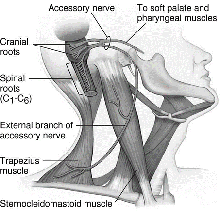
Accessory
(XI) Nerve (Motor)
• Originates in the anterior gray horn of the first 5
cervical segments of the spinal cord
• Emerges laterally from the cord and then ascends through
the foramen magnum into the cranial cavity
• It then arches inferiorly to leave the jugular foramen
• Function: mediates movement of head and pectoral girdle
Hypoglossal
(XII) Nerve (Motor)
• Originates in the medulla
• passes through the hypoglossal canal, and supplies muscles
of the tongue
• Function: Movement of tongue during speech and swallowing

Spinal Cord
• Contribute to homeostasis by providing quick, reflexive
responses to many stimuli
• Pathway for sensory input to the brain and motor output
from the brain

Protective
Structures of Spinal Cord
• Two types of connective tissue coverings
• Bony vertebrae
• Tough, connective tissue meninges—plus a cushion of CSF
• Surround and protect the delicate nervous tissue of the
spinal cord

Protective
Structures - Vertebral Column
• Spinal cord is located within the vertebral canal of the
vertebral column
• Vertebral foramina of all the vertebrae, stacked one on
top of the other, form the vertebral canal
• The surrounding vertebrae provide a sturdy shelter for the
enclosed spinal cord

Meninges
• Three connective tissue coverings encircle the spinal cord
and brain
• Continuous with the cranial meninges

Dura mater
• Superficial spinal meninges
• Composed of dense, irregular connective tissue
• Epidural space - Between dura mater & wall of
vertebral canal
Arachnoid mater
• Middle a vascular covering
• Continuous with arachnoid mater of the brain
• Subdural space – Between dura mater and the arachnoid
Pia mater
• Innermost meninge - Thin transparent connective tissue
• Blood vessels – Supply oxygen and nutrients
• Subarachnoid space: Between arachanoid and pia mater (CSF)
External
Anatomy of Spinal Cord
• Roughly cylindrical (flattened slightly anteriorly &
posteriorly)
• Extends from the medulla oblongata to the superior border
of the second lumbar vertebra
• In newborn, extends to the third or fourth lumbar vertebra
• Two conspicuous of spinal cord
– Superior enlargement – cervical enlargement C4 - T1
– Inferior enlargement – lumbar enlargement T9 - T12

• Inferior to the lumbar enlargement, the spinal cord
terminates as a tapering, conical structure - conus medullaris
• Ends at the level of the intervertebral disc between L1-L2
Filum terminale
– Arise from the conus medullaris
– Extension of pia mater that extends inferiorly
– Blends with the arachnoid mater and dura mater
– Anchors the spinal cord to the coccyx
Spinal
Nerves
• Paths of communication between the spinal cord and
specific regions of the body
• 31 pairs of spinal nerves emerge at regular intervals from
intervertebral foramina
• Each pair of spinal nerves arise from a spinal segment
• Roots of these spinal nerves angle inferiorly in the
vertebral canal from the end of the spinal cord
• Branched nerves connect the CNS to the sensory receptors,
muscles, and glands in all parts of the body
There are:
• 8 pairs cervical
nerves (C1–C8)
• 12 pairs of
thoracic nerves (T1–T12)
• 5 pairs of lumbar
nerves (L1–L5)
• 5 pairs of sacral
nerves (S1–S5)
• 1 pair of
coccygeal nerves (co1)

• Roots -Two
bundles of axons
• Rootlets -
connect each spinal nerve to a segment of the cord by even smaller bundles of
axons

Posterior (dorsal)
root and rootlets
• Contain only sensory axons
• Conduct nerve impulses from sensory receptors in the skin,
muscles, and internal organs into the CNS
• Each posterior root has a swelling - posterior (dorsal)
root ganglion
Anterior (ventral)
root and rootlets
• Contain axons of motor neurons
• Conduct nerve impulses from the CNS to effectors (muscles
and glands)

Internal
Anatomy of Spinal Cord

• Spinal cord reveals regions of white matter that surround
an inner core of gray matter
• Two grooves penetrate the white matter
• Divide spinal cord into right and left sides
• Anterior median fissure
• Posterior median sulcus
• Gray matter of the spinal cord is shaped like the letter H
or a butterfly
• Gray commissure forms the crossbar of the H
• Small space in the center of the gray commissure - Central
canal
• Extends the entire length of spinal cord (filled with CSF)
Gray Matter
of Spinal Cord
• Gray matter on each side of the spinal cord is subdivided
into regions called horns
Posterior (dorsal) gray
horns
• Contain cell bodies and axons of interneurons as well as
axons of incoming sensory neurons
Anterior (ventral) gray
horns
• Contain somatic motor nuclei
• Provide nerve impulses for contraction of skeletal muscles
Lateral gray horns
• Present only in thoracic & upper lumbar segments of
spinal cord
• Contain autonomic motor nuclei
White
matter of Spinal Cord
• Organized into regions
• The anterior and posterior gray horns divide the white
matter on each side into three broad areas:
– Anterior (ventral) white columns
– Posterior (dorsal) white columns
– Lateral white columns
• Each columns – bundles of axons
Processing of sensory
input and motor output by the spinal cord

Physiology
of Spinal Cord
• The spinal cord has two principal functions in maintaining
homeostasis:
– Nerve impulse propagation
– Integration of information
• White matter tracts are highways for nerve impulse
propagation
• Sensory input travels along these tracts toward the brain
• Motor output travels from the brain along these tracts
toward skeletal muscles and other effector tissues
• Gray matter of the spinal cord receives and integrates
incoming and outgoing information
Reflexes and
Reflex Arc
• Spinal cord promotes homeostasis is by serving as an
integrating center for some reflexes
• Reflex is a fast,
automatic, unplanned sequence of actions that occurs in response to a
particular stimulus
• Some reflexes – inborn: pulling hand away from a hot
surface feel
• Other reflexes are learned or acquired: driving

Reflex Arc

• The pathway followed by nerve impulses that produce a
reflex is a reflex arc
• Five functional components
– Sensory receptor
– Sensory neuron
– Integrating center
– Motor neuron
– Effector
Reflexes
Types and Examples
• Reflexes may be spinal or cranial and somatic or autonomic
• Somatic spinal reflexes include the stretch reflex, the
tendon reflex, the flexor (withdrawal) reflex & crossed extensor reflex
• All exhibit reciprocal innervation
• Monosynaptic reflex
arc: Consists of one sensory & one motor neuron (stretch reflex –
patellar reflex, is an example)
• Polysynaptic reflex
arc: Contains sensory neurons, interneurons, and motor neurons
Stretch Reflex

Tendon Reflex

Autonomic
Nervous System
• Contributes to homeostasis by responding to subconscious
visceral sensations
• Operates without conscious control
• Peripheral nervous system (PNS) includes:
– Cranial and spinal nerves
– Divided into: SNS, ANS & ENS
• Structurally, ANS includes:
– Autonomic sensory neurons
– Integrating centers in the CNS
– Autonomic motor neurons

Division of
ANS
The output (motor) part of the ANS has two divisions:
Sympathetic
Parasympathetic

ANS &
Visceral Activities
• Autonomic motor neurons regulate visceral activities by
either increasing (exciting) or decreasing (inhibiting) ongoing activities in
their effector tissues (cardiac muscle, smooth muscle, and glands)


Motor
Neuron Pathways


Anatomical
Components of ANS


Preganglionic
Neurons


Autonomic
Ganglia
Sympathetic ganglia
• Sympathetic trunk ganglia: superior, middle, and inferior
cervical ganglia
• Prevertebral ganglia: celiac, superior mesenteric,
inferior mesenteric, aorticorenal, renal ganglion
Parasympathetic
ganglia
• Terminal ganglia (intramural ganglia): Ciliary ganglion,
pterygopalatine ganglion, submandibular ganglion, and otic ganglion
Parasympathetic
Division
• Axons of sympathetic preganglionic neurons pass to
sympathetic trunk ganglia
• They may connect with postganglionic neurons in different
ways

Sympathetic
Division

Autonomic
Plexuses
• Tangled networks of axons of both sympathetic and
parasympathetic neurons
• Many lie along major arteries
• They are:
– Cardiac plexus
– Pulmonary plexus
– Celiac plexus
– Superior mesenteric plexus
– Inferior mesenteric plexus
– Hypogastric plexus
– Renal plexus

ANS -
Neurotransmission

Physiology
of ANS

Autonomic
Reflex
• Response that occur when nerve impulses pass through an
autonomic reflux arc
Regulate controlled conditions in the body like
Blood pressure –
by adjusting - heart rate, contraction of ventricles, diameter of blood vessels
Digestion – by
adjusting motility and muscle tone of GIT
Defecation and
Urination – by regulating opening and closing of sphincters
Components
of autonomic reflex
Receptor
• In the distal end of sensory neuron
• Responds to stimulus and produce nerve impulse
• Associated with interoceptor
Sensory neurons
• Conduct nerve impulse from receptor to CNS
Integrating centre
• Interneurons in CNS relay signals from sensory to motor
neuron
• Located in hypothalamus and brain stem
Motor neurons
• Carry impulse out of CNS
• Preganglionic neuron and post ganglionic neuron
Effector
• Smooth muscles, cardiac muscles, and glands
Autonomic
Control by Higher Centers
Hypothalamus
• Major control and integration center of the ANS
• Receives sensory input related to visceral functions,
olfaction, gustation, changes in temperature, osmolarity & levels of
various substances in blood
• Also receives input related to emotions from the limbic
system
• Output from the hypothalamus influences autonomic centers
in both:
– Brain stem (such as the cardiovascular, salivation
swallowing, and vomiting centers)
– Spinal cord (such as the defecation and urination reflex
centers in the sacral spinal cord)

• The posterior and lateral parts of the hypothalamus
control the sympathetic division
• The anterior and medial parts of the hypothalamus control
the parasympathetic division
Summary
• Structures of nervous system - the brain, cranial nerves
(12 pairs) and their branches, the spinal cord, spinal nerves (31 pairs) and
their branches, ganglia, enteric plexuses, and sensory receptors
• Functions- Homeostasis and integrates all body activities
by sensing changes, interpreting them and reacting to them
• Nervous tissue consists of neuron and neuroglia
• Parts of neuron – Dendrite, Axon & cell body
• Neuroglia support, nurture, and protect neurons and
maintain the interstitial fluid
• White matter - aggregates of myelinated axons
• Gray matter - cell bodies, dendrites, and axon terminals
of neurons, unmyelinated axons, and neuroglia
• Plasma membranes of presynaptic and postsynaptic neurons -
separated by the synaptic cleft
• Release of neurotransmitter, postsynaptic neuron receives
the chemical signal, produces a postsynaptic potential
• Postsynaptic potential may be a depolarization or a
hyperpolarization, depends on the neurotransmitter released
• CNS consists of the brain and spinal cord
• PNS consists of somatic nervous system (SNS), autonomic
nervous system (ANS), and enteric nervous system (ENS)
• SNS - Sensory neurons, conduct impulses from somatic and
special sense receptors to the CNS; motor neurons from the CNS to skeletal
muscles
• ANS - Sensory neurons from visceral organs and motor
neurons; convey impulses from the CNS to smooth muscle tissue, cardiac muscle
tissue, and glands
• ENS consists of neurons in enteric plexuses in the
gastrointestinal (GI) tract
• Major parts of the brain are the brain stem, cerebellum,
diencephalon, and cerebrum
• Brain is protected by cranial bones and the cranial
meninges
• Blood flow to the brain is mainly via the internal carotid
and vertebral arteries
• Blood–brain barrier (BBB) causes different substances to
move between the blood and the brain tissue
• Cerebrospinal fluid (CSF) is formed in the choroid
plexuses and circulates through various ventricles
• CSF provides mechanical protection, chemical protection,
and circulation of nutrients
• Brain stem consists of medulla oblongata, pons and mid
brain
• Medulla oblongata- continuous with spinal cord, has vital
center like respiratory, cardiovascular, vomiting, deglutition
• Pons - lies superior to the medulla and anterior to the
cerebellum; Signals for voluntary movements from motor areas of the cerebral
cortex are relayed
• Mid brain - Extends from the pons to the diencephalon; has
centers for various reflexes
• Reticular formation-Netlike region of interspersed gray
and white matter; extends through the brain stem
• Cerebellum - occupies the inferior and posterior aspects
of the cranial cavity
• The anterior lobe and posterior lobe govern subconscious
aspects of skeletal muscle movements
• Flocculonodular lobe on the inferior surface contributes
to equilibrium and balance
• Diencephalon surrounds the third ventricle and consists of
the thalamus, hypothalamus, and epithalamus
• Thalamus is superior to the midbrain and contains nuclei
• Thalamus serve as relay stations for most sensory input to
the cereberal cortex
• Hypothalamus is inferior to the thalamus
• It controls the ANS, produces hormones, and regulates
emotional and behavioral patterns; controls body temperature by serving as the
body’s thermostat
• Circumventricular organs (CVOs) can monitor chemical changes
in the blood because they lack the blood–brain barrier
• Cerebrum is the largest part of the brain
• Its cortex contains gyri (convolutions), fissures, and
Sulci
• Cerebral hemispheres are divided into four lobes: frontal,
parietal, temporal, and occipital
• White matter of the cerebrum is deep to the cortex
• It consists primarily of myelinated axons extending to
other regions as association,commissural,
and projection fibers
• Basal ganglia are several groups of nuclei in each
cerebral hemisphere
• Help initiate and terminate movements, suppress unwanted
movements, and regulate muscle tone
• Limbic system encircles the upper part of the brain stem
and the corpus callosum
• It functions in emotional aspects of behavior and memory
• Sensory areas of the cerebral cortex allow perception of
sensory information
• The motor areas control the execution of voluntary
movements
• The association areas are concerned with more complex
integrative functions such as memory, personality traits, and intelligence
• Brain waves generated by the cerebral cortex are recorded
from the surface of the head in an electroencephalogram (EEG)
• EEG may be used to diagnose epilepsy, infections, and
tumors
• Twelve pairs of cranial nerves originate from the nose,
eyes, inner ear, brain stem, and spinal cord
• Three cranial nerves (I, II, and VIII) carry axons of
sensory neurons - sensory nerves
• Five cranial nerves (III, IV, VI, XI, and XII) contain
only axons of motor neurons as they leave the brain stem - motor nerves
• Four cranial nerves (V, VII, IX, and X) are mixed nerves
because they contain axons of both sensory and motor neurons
• The spinal cord is protected by the vertebral column, the
meninges, cerebrospinal fluid, and denticulate ligaments
• 3 meninges (dura mater, arachnoid mater, and pia mater)
are coverings that run continuously around the spinal cord and brain
• 31 pair of spinal nerves connect to each segment of the
spinal cord by two roots
• There are 8 pairs of cervical, 12 pairs of thoracic, 5
pairs of lumbar, 5 pairs of sacral, and 1 pair of coccygeal nerves
• A reflex is a fast, predictable sequence of involuntary
actions, such as muscle contractions or glandular secretions, which occurs in
response to certain changes in the environment
• The pathway followed by nerve impulses that produce a
reflex is a reflex arc
• Components of a reflex arc - sensory receptor, sensory
neuron, integrating center, motor
• ANS comprises of sympathetic and parasympathetic division
• Sympathetic division favors body functions that can
support vigorous physical activity and rapid production of ATP (fight-or flight
response)
• The parasympathetic division regulates activities that
conserve and restore body energy
• Autonomic ganglia comprises of sympathetic and
parasympathetic ganglia
• Sympathetic ganglia include sympathetic trunk ganglia and
pre vertebral ganglia
• Parasympathetic ganglia are known as terminal ganglia
• Hypothalamus is the major control and integration center
of the ANS
• It is connected to both the sympathetic and the
parasympathetic divisions


0 Comments